Thursday, June 30, 2022

Enamel Loss

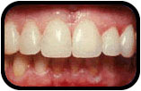
The high mineral content of enamel, which makes this tissue the hardest in the human body, also makes it demineralize in a process that often occurs as dental caries, otherwise known as cavities. Demineralization occurs for several reasons, but the most important cause of tooth decay is the ingestion of fermentable carbohydrates.[citation needed] Tooth cavities are caused when acids dissolve tooth enamel: Enamel is also lost through tooth wear and enamel fractures.

Ca10(PO4)6(OH)2(s) + 8H+(aq) → 10Ca2+(aq) + 6HPO42−(aq) + 2H2O(l)

Sugars and acids from candies, soft drinks, and fruit juices play a significant role in tooth decay, and consequently in enamel destruction. The mouth contains a great number and variety of bacteria, and when sucrose, the most common of sugars, coats the surface of the mouth, some intraoral bacteria interact with it and form lactic acid, which decreases the pH in the mouth. The critical pH for tooth enamel is generally accepted to be pH 5.5. When acids are present and the critical pH is reached, the hydroxyapatite crystallites of enamel demineralize, allowing for greater bacterial invasion deeper into the tooth. The most important bacterium involved with tooth decay is Streptococcus mutans, but the number and type of bacteria varies with the progress of tooth destruction.

Furthermore, tooth morphology dictates that the most common site for the initiation of dental caries is in the deep grooves, pits, and fissures of enamel. This is expected because these locations are impossible to reach with a toothbrush and allow for bacteria to reside there. When demineralization of enamel occurs, a dentist can use a sharp instrument, such as a dental explorer, and "feel a stick" at the location of the decay. As enamel continues to become less mineralized and is unable to prevent the encroachment of bacteria, the underlying dentin becomes affected as well. When dentin, which normally supports enamel, is destroyed by a physiologic condition or by decay, enamel is unable to compensate for its brittleness and breaks away from the tooth easily.

The extent to which tooth decay is likely, known as cariogenicity, depends on factors such as how long the sugar remains in the mouth. Contrary to common belief, it is not the amount of sugar ingested but the frequency of sugar ingestion that is the most important factor in the causation of tooth decay. When the pH in the mouth initially decreases from the ingestion of sugars, the enamel is demineralized and left vulnerable for about 30 minutes. Eating a greater quantity of sugar in one sitting does not increase the time of demineralization. Similarly, eating a lesser quantity of sugar in one sitting does not decrease the time of demineralization. Thus, eating a great quantity of sugar at one time in the day is less detrimental than is a very small quantity ingested in many intervals throughout the day. For example, in terms of oral health, it is better to eat a single dessert at dinner time than to snack on a bag of candy throughout the day.

In addition to bacterial invasion, enamel is also susceptible to other destructive forces. Bruxism, also known as clenching of or grinding on teeth, destroys enamel very quickly. The wear rate of enamel, called attrition, is 8 micrometers a year from normal factors. A common misconception is that enamel wears away mostly from chewing, but actually teeth rarely touch during chewing. Furthermore, normal tooth contact is compensated physiologically by the periodontal ligaments (pdl) and the arrangement of dental occlusion. The truly destructive forces are the parafunctional movements, as found in bruxism, which can cause irreversible damage to the enamel.

Other nonbacterial processes of enamel destruction include abrasion (involving foreign elements, such as toothbrushes), erosion (involving chemical processes, such as dissolving by soft drinks or lemon and other juices), and possibly abfraction (involving compressive and tensile forces).

Though enamel is described as tough, it has a similar brittleness to glass, making it, unlike other natural crack-resistant laminate structures such as shell and nacre, vulnerable to fracture. In spite of this it can withstand bite forces as high as 1,000 N many times a day during chewing. This resistance is due in part to the microstructure of enamel which contains enamel tufts that stabilize such fractures at the dentinoenamel junction. The configuration of the tooth also acts to reduce the tensile stresses that cause fractures during biting.

Gastroesophageal reflux disease can also lead to enamel loss, as acid refluxes up the esophagus and into the mouth, occurring most during overnight sleep.

Read more, here.

707-453-1776
1001 Nut Tree Rd, Ste 230
Vacaville, CA 95687
donnathedentist@gmail.com

Monday, June 27, 2022

Can Tooth Enamel Grow Back? (How To Prevent Enamel Erosion)


To prevent enamel erosion - you can take the same exact steps that you would to remineralize your teeth. This video talks about the 8 tips to restrengthen your enamel (remineralize your teeth) and prevent enamel erosion!

707-453-1776
1001 Nut Tree Rd, Ste 230
Vacaville, CA 95687
donnathedentist@gmail.com

Friday, June 24, 2022

Development of Enamel Formation


Enamel formation is part of the overall process of tooth development. Under a microscope, different cellular aggregations are identifiable within the tissues of a developing tooth, including structures known as the enamel organ, dental lamina, and dental papilla. The generally recognized stages of tooth development are the bud stage, cap stage, bell stage, and crown, or calcification, stage. Enamel formation is first seen in the crown stage.

Amelogenesis, or enamel formation, occurs after the first establishment of dentin, via cells known as ameloblasts. Human enamel forms at a rate of around 4 μm per day, beginning at the future location of cusps, around the third or fourth month of pregnancy. As in all human processes, the creation of enamel is complex, but can generally be divided into two stages. The first stage, called the secretory stage, involves proteins and an organic matrix forming a partially mineralized enamel. The second stage, called the maturation stage, completes enamel mineralization.

In the secretory stage, ameloblasts are polarized columnar cells. In the rough endoplasmic reticulum of these cells, enamel proteins are released into the surrounding area and contribute to what is known as the enamel matrix, which is then partially mineralized by the enzyme alkaline phosphatase. When this first layer is formed, the ameloblasts move away from the dentin, allowing for the development of Tomes' processes at the apical pole of the cell. Enamel formation continues around the adjoining ameloblasts, resulting in a walled area, or pit, that houses a Tomes' process, and also around the end of each Tomes' process, resulting in a deposition of enamel matrix inside of each pit. The matrix within the pit will eventually become an enamel rod, and the walls will eventually become interrod enamel. The only distinguishing factor between the two is the orientation of the calcium phosphate crystallites.

In the maturation stage, the ameloblasts transport substances used in the formation of enamel. Histologically, the most notable aspect of this phase is that these cells become striated, or have a ruffled border. These signs demonstrate that the ameloblasts have changed their function from production, as in the secretory stage, to transportation. Proteins used for the final mineralization process compose most of the transported material. The noteworthy proteins involved are amelogenins, ameloblastins, enamelins, and tuftelins. How these proteins are secreted into the enamel structure is still unknown; other proteins, such as the Wnt signaling components BCL9 and Pygopus, have been implicated in this process. During this process, amelogenins and ameloblastins are removed after use, leaving enamelins and tuftelin in the enamel. By the end of this stage, the enamel has completed its mineralization.

At some point before the tooth erupts into the mouth, but after the maturation stage, the ameloblasts are broken down. Consequently, enamel, unlike many other tissues of the body, has no way to regenerate itself. After destruction of enamel from decay or injury, neither the body nor a dentist can restore the enamel tissue. Enamel can be affected further by non-pathologic processes.

Enamel is covered by various structures in relation to the development of tooth:
  • Nasmyth membrane or enamel cuticle, structure of embryological origin is composed of keratin which gives rise to the enamel organ.
  • Acquired pellicle, structure acquired after tooth eruption is composed of food debris, calculus, dental plaque (organic film).

Read more, here.

707-453-1776
1001 Nut Tree Rd, Ste 230
Vacaville, CA 95687
donnathedentist@gmail.com


Tuesday, June 21, 2022

What is Tooth Enamel?

Tooth enamel is one of the four major tissues that make up the tooth in humans and many other animals, including some species of fish. It makes up the normally visible part of the tooth, covering the crown. The other major tissues are dentin, cementum, and dental pulp. It is a very hard, white to off-white, highly mineralised substance that acts as a barrier to protect the tooth but can become susceptible to degradation, especially by acids from food and drink. Calcium hardens the tooth enamel. In rare circumstances enamel fails to form, leaving the underlying dentin exposed on the surface.

Enamel is the hardest substance in the human body and contains the highest percentage of minerals (at 96%), with water and organic material composing the rest. The primary mineral is hydroxyapatite, which is a crystalline calcium phosphate. Enamel is formed on the tooth while the tooth develops within the jaw bone before it erupts into the mouth. Once fully formed, enamel does not contain blood vessels or nerves, and is not made of cells. Remineralisation of teeth can repair damage to the tooth to a certain degree but damage beyond that cannot be repaired by the body. The maintenance and repair of human tooth enamel is one of the primary concerns of dentistry.

In humans, enamel varies in thickness over the surface of the tooth, often thickest at the cusp, up to 2.5 mm, and thinnest at its border with the cementum at the cementoenamel junction (CEJ).

The normal color of enamel varies from light yellow to grayish (bluish) white. At the edges of teeth where there is no dentin underlying the enamel, the color sometimes has a slightly blue or translucent off-white tone, easily observable on the upper incisors. Since enamel is semitranslucent, the color of dentin and any material underneath the enamel strongly affects the appearance of a tooth. The enamel on primary teeth has a more opaque crystalline form and thus appears whiter than on permanent teeth.

The large amount of mineral in enamel accounts not only for its strength but also for its brittleness. Tooth enamel ranks 5 on Mohs hardness scale (between steel and titanium) and has a Young's modulus of 83 GPa. Dentin, less mineralized and less brittle, 3–4 in hardness, compensates for enamel and is necessary as a support. On radiographs, the differences in the mineralization of different portions of the tooth and surrounding periodontium can be noted; enamel appears lighter than dentin or pulp since it is denser than both and more radiopaque.

Enamel does not contain collagen, as found in other hard tissues such as dentin and bone, but it does contain two unique classes of proteins: amelogenins and enamelins. While the role of these proteins is not fully understood, it is believed that they aid in the development of enamel by serving as a framework for minerals to form on, among other functions. Once it is mature, enamel is almost totally without the softer organic matter. Enamel is avascular and has no nerve supply within it and is not renewed, however, it is not a static tissue as it can undergo mineralization changes.

Read more, here.

707-453-1776
1001 Nut Tree Rd, Ste 230
Vacaville, CA 95687
donnathedentist@gmail.com


Saturday, June 18, 2022

Tooth Decay (Cavity) Explained | Cavities vs Fillings vs Dental Caries


What is tooth decay?  What is a cavity?  Is dental caries a thing?  Where do dental fillings come in?  Let's talk!
Cavities in teeth are caused by the acid byproducts of plaque biofilm and the foods/drinks that we consume. Thankfully, they’re preventable with good daily dental care and regular checkups. The symptoms of cavities include things like sensitivity, pain, rough edges, and discoloration.

Fillings are the best treatment for cavities, but sometimes a crown or root canal is necessary, especially if complications like abscesses develop. If you catch cavities in the earliest stages, you can prevent the bacteria from eroding totally through your enamel. See your dentist at least every six months to help reduce your risk of tooth decay and intercept it when it’s small.

707-453-1776
1001 Nut Tree Rd, Ste 230
Vacaville, CA 95687
donnathedentist@gmail.com

Wednesday, June 15, 2022


Four things are required for caries to form: a tooth surface (enamel or dentin), caries-causing bacteria, fermentable carbohydrates (such as sucrose), and time. This involves adherence of food to the teeth and acid creation by the bacteria that makes up the dental plaque. However, these four criteria are not always enough to cause the disease and a sheltered environment promoting development of a cariogenic biofilm is required. The caries disease process does not have an inevitable outcome, and different individuals will be susceptible to different degrees depending on the shape of their teeth, oral hygiene habits, and the buffering capacity of their saliva. Dental caries can occur on any surface of a tooth that is exposed to the oral cavity, but not the structures that are retained within the bone.

Tooth decay is caused by biofilm (dental plaque) lying on the teeth and maturing to become cariogenic (causing decay). Certain bacteria in the biofilm produce acid in the presence of fermentable carbohydrates such as sucrose, fructose, and glucose.

Caries occur more often in people from the lower end of the socio-economic scale than people from the upper end of the socio-economic scale.

Read more, here.
707-453-1776
1001 Nut Tree Rd, Ste 230
Vacaville, CA 95687
donnathedentist@gmail.com

Sunday, June 12, 2022

Signs and Symptoms of Tooth Decay


A person experiencing caries may not be aware of the disease. The earliest sign of a new carious lesion is the appearance of a chalky white spot on the surface of the tooth, indicating an area of demineralization of enamel. This is referred to as a white spot lesion, an incipient carious lesion or a "micro-cavity". As the lesion continues to demineralize, it can turn brown but will eventually turn into a cavitation ("cavity"). Before the cavity forms, the process is reversible, but once a cavity forms, the lost tooth structure cannot be regenerated. A lesion that appears dark brown and shiny suggests dental caries were once present but the demineralization process has stopped, leaving a stain. Active decay is lighter in color and dull in appearance.

As the enamel and dentin are destroyed, the cavity becomes more noticeable. The affected areas of the tooth change color and become soft to the touch. Once the decay passes through the enamel, the dentinal tubules, which have passages to the nerve of the tooth, become exposed, resulting in pain that can be transient, temporarily worsening with exposure to heat, cold, or sweet foods and drinks. A tooth weakened by extensive internal decay can sometimes suddenly fracture under normal chewing forces. When the decay has progressed enough to allow the bacteria to overwhelm the pulp tissue in the center of the tooth, a toothache can result and the pain will become more constant. Death of the pulp tissue and infection are common consequences. The tooth will no longer be sensitive to hot or cold but can be very tender to pressure.

Dental caries can also cause bad breath and foul tastes. In highly progressed cases, an infection can spread from the tooth to the surrounding soft tissues. Complications such as cavernous sinus thrombosis and Ludwig angina can be life-threatening.

Read more, here.

707-453-1776
1001 Nut Tree Rd, Ste 230
Vacaville, CA 95687
donnathedentist@gmail.com

Thursday, June 9, 2022

Toothpaste Hacks (1 Bad Hack & 5 Good Hacks)


Let's talk about different "toothpaste beauty hacks" and whether or not they are worth your while!  (PS - toothpaste on pimples is a big NO!)

Putting toothpaste on pimples won’t help as much as you may hope it does. In reality, it could just irritate your face and make things worse. The better option is to promote healthy skin with a good oral hygiene and face care routine to limit the bacteria and oils responsible for causing breakouts. But there are still plenty of other useful lifehacks you can get out of using toothpaste around your house. Just try not to use it on your face!

707-453-1776
1001 Nut Tree Rd, Ste 230
Vacaville, CA 95687
donnathedentist@gmail.com

Monday, June 6, 2022

Tooth Decay


Tooth decay, also known as cavities or caries, is the breakdown of teeth due to acids produced by bacteria. The cavities may be a number of different colors from yellow to black. Symptoms may include pain and difficulty with eating. Complications may include inflammation of the tissue around the tooth, tooth loss and infection or abscess formation.

The cause of cavities is acid from bacteria dissolving the hard tissues of the teeth (enamel, dentin and cementum). The acid is produced by the bacteria when they break down food debris or sugar on the tooth surface. Simple sugars in food are these bacteria's primary energy source and thus a diet high in simple sugar is a risk factor. If mineral breakdown is greater than build up from sources such as saliva, caries results. Risk factors include conditions that result in less saliva such as: diabetes mellitus, Sjögren syndrome and some medications. Medications that decrease saliva production include antihistamines and antidepressants. Dental caries are also associated with poverty, poor cleaning of the mouth, and receding gums resulting in exposure of the roots of the teeth.

Prevention of dental caries includes regular cleaning of the teeth, a diet low in sugar, and small amounts of fluoride. Brushing one's teeth twice per day and flossing between the teeth once a day is recommended. Fluoride may be acquired from water, salt or toothpaste among other sources. Treating a mother's dental caries may decrease the risk in her children by decreasing the number of certain bacteria she may spread to them. Screening can result in earlier detection. Depending on the extent of destruction, various treatments can be used to restore the tooth to proper function or the tooth may be removed. There is no known method to grow back large amounts of tooth. The availability of treatment is often poor in the developing world. Paracetamol (acetaminophen) or ibuprofen may be taken for pain.

Worldwide, approximately 3.6 billion people (48% of the population) have dental caries in their permanent teeth as of 2016. The World Health Organization estimates that nearly all adults have dental caries at some point in time. In baby teeth it affects about 620 million people or 9% of the population. They have become more common in both children and adults in recent years. The disease is most common in the developed world due to greater simple sugar consumption and less common in the developing world. Caries is Latin for "rottenness".

Read more, here.

707-453-1776
1001 Nut Tree Rd, Ste 230
Vacaville, CA 95687
donnathedentist@gmail.com

Friday, June 3, 2022

Dental Radiography


Dental radiographs are commonly called X-rays. Dentists use radiographs for many reasons: to find hidden dental structures, malignant or benign masses, bone loss, and cavities.

A radiographic image is formed by a controlled burst of X-ray radiation which penetrates oral structures at different levels, depending on varying anatomical densities, before striking the film or sensor. Teeth appear lighter because less radiation penetrates them to reach the film. Dental caries, infections and other changes in the bone density, and the periodontal ligament, appear darker because X-rays readily penetrate these less dense structures. Dental restorations (fillings, crowns) may appear lighter or darker, depending on the density of the material.

The dosage of X-ray radiation received by a dental patient is typically small (around 0.150 mSv for a full mouth series[1]), equivalent to a few days' worth of background environmental radiation exposure, or similar to the dose received during a cross-country airplane flight (concentrated into one short burst aimed at a small area). Incidental exposure is further reduced by the use of a lead shield, lead apron, sometimes with a lead thyroid collar. Technician exposure is reduced by stepping out of the room, or behind adequate shielding material, when the X-ray source is activated.

Once photographic film has been exposed to X-ray radiation, it needs to be developed, traditionally using a process where the film is exposed to a series of chemicals in a dark room, as the films are sensitive to normal light. This can be a time-consuming process, and incorrect exposures or mistakes in the development process can necessitate retakes, exposing the patient to additional radiation. Digital X-rays, which replace the film with an electronic sensor, address some of these issues, and are becoming widely used in dentistry as the technology evolves. They may require less radiation and are processed much more quickly than conventional radiographic films, often instantly viewable on a computer. However digital sensors are extremely costly and have historically had poor resolution, though this is much improved in modern sensors.

It is possible for both tooth decay and periodontal disease to be missed during a clinical exam, and radiographic evaluation of the dental and periodontal tissues is a critical segment of the comprehensive oral examination. The photographic montage at right depicts a situation in which extensive decay had been overlooked by a number of dentists prior to radiographic evaluation.

Read more, here.

707-453-1776
1001 Nut Tree Rd, Ste 230
Vacaville, CA 95687
donnathedentist@gmail.com