Let's talk about why mouth breathing is AWFUL for your dental health!
Typically, your mouth needs plenty of saliva to stay healthy. An adequate saliva flow helps to limit the accumulation of bacteria, dry tissues, and even odors. When someone is always breathing through their mouth instead of their nose—say for the occasional hard workout at the gym—it dries out your saliva quicker than it can replenish itself.
The more you breathe out your mouth, the drier your mouth becomes. And the drier your mouth is, the higher your chances are for:
• Cavities
• Halitosis
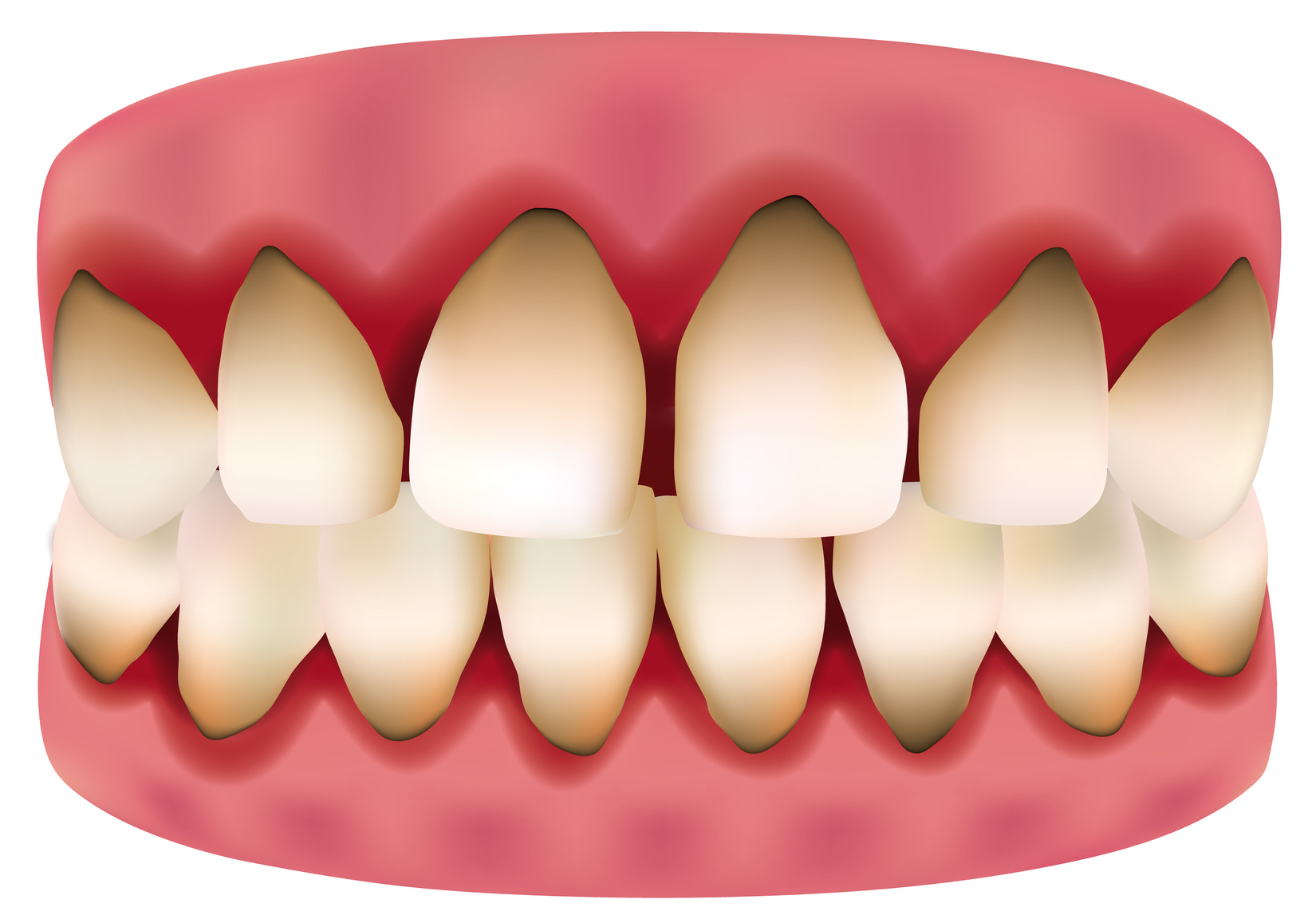
• Gum disease
• Dry, cracked lips
In fact, in dentistry we purposely try to treat and manage dry mouth, because it’s that bad for your tooth enamel. Without saliva coating your teeth throughout the day, your bacterial and acid levels skyrocket. Chronic mouth breathing can—and almost always will—lead to an uptick in dental diseases. You could almost equate mouth breathing to being as bad on your teeth and gums as what we see with cancer patients undergoing radiation therapy. It’s a huge factor in your dental wellness, not just a minor knit-picky issue.